
Alberto Jiménez Schuhmacher highlights on Onda Cero the potential of the eclipse to awaken new scientific vocations
August 14, 2026
A drug uses glucose to slow the spread of cancer in mice
August 19, 2026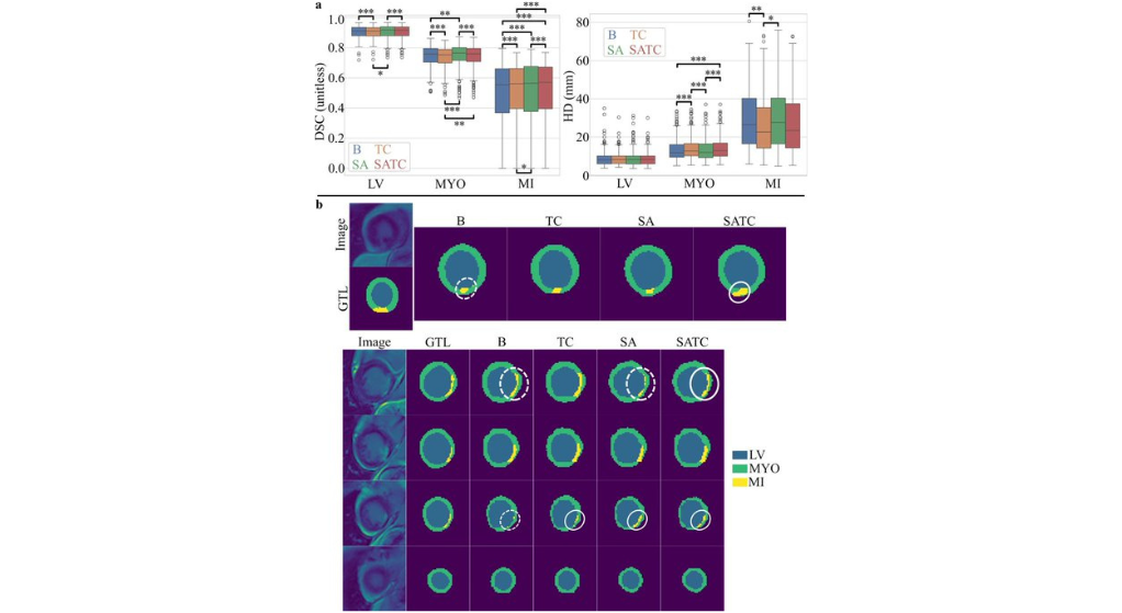
Researchers from the Aragon Health Research Institute, the University of Zaragoza, and the I3A are developing new computational strategies to obtain more reliable maps of the heart and facilitate the analysis of medical images in different clinical settings.
Obtaining accurate maps of the heart from cardiac magnetic resonance imaging remains a challenge, especially when the images come from different hospitals, acquisition systems, or patient populations. Improving the reliability of these analyses is key to advancing diagnosis and personalized treatment planning.
A study developed by researchers from Aragón Health Research Institute (IIS Aragón), the University of Zaragoza and the Engineering Research Institute of Aragon (I3A) has evaluated new strategies based on artificial intelligence to optimize the three-dimensional segmentation of cardiac magnetic resonance imaging.
The work, published in the journal Annals of Biomedical Engineering, analyzes the impact of combining two computational techniques: the data augmentation using synthetic images and topological correction, applied to studies from multiple centers, technology providers, image contrasts and tissue types.
The results show that combining both tools improves the quality of cardiac segmentations. On the one hand, enlarging synthetic images increases the match with the reference labels used by specialists; on the other, topological correction reduces structural errors and particularly improves the identification of areas damaged by a myocardial infarction.
Automatic segmentation of medical images allows for the delineation of cardiac structures and the extraction of relevant information for study. However, differences between MRI equipment, clinical protocols, and patient characteristics can affect the performance of the algorithms. This study demonstrates that incorporating data enhancement and structural control strategies can contribute to the development of more robust tools applicable in real-world clinical settings.
The authors of the work, Ricardo M. Rosales, Manuel Doblaré and Esther PueyoResearchers from IIS Aragón, I3A and the University of Zaragoza point out that these advances can promote a more consistent computational analysis of cardiac imaging, facilitating its application in different health centers and population groups.
The research represents a new advance in the development of tools to support cardiovascular diagnosis, promoting the use of artificial intelligence to improve the accuracy and reproducibility of medical imaging studies.
Study reference: 3D Segmentation of Multi-Contrast Cardiac Magnetic Resonance Images with Topological Correction and Synthetic Data Augmentation.
Authors: Ricardo M. Rosales, Manuel Doblaré and Esther Pueyo.